-
Type of microscope (Light Microscope, Electron Microscope)Biological Science 2020. 10. 26. 12:22
Type of microscope
Yes. It's easy to understand that cells are small, so you have to zoom in. By the way, why do we have to learn these kinds of machines while studying biology? It is to understand each of the pros and cons and to understand what the appropriate microscope is, depending on what you want to observe, and to better interpret the various microscopic photographs you will see in the future.
The eggs of long nerve cells up to one meter long and the frog in the diameter of several millimeters can be seen with the naked eye without the help of a microscope. However, most cells are 1 to 100 mm in size and can be examined through optical microscopes. And an electron microscope is needed to observe smaller cell organs. This is well illustrated in the picture below. Let's look at each of the characteristics of the optical microscope and the electron microscope in the following paragraphs.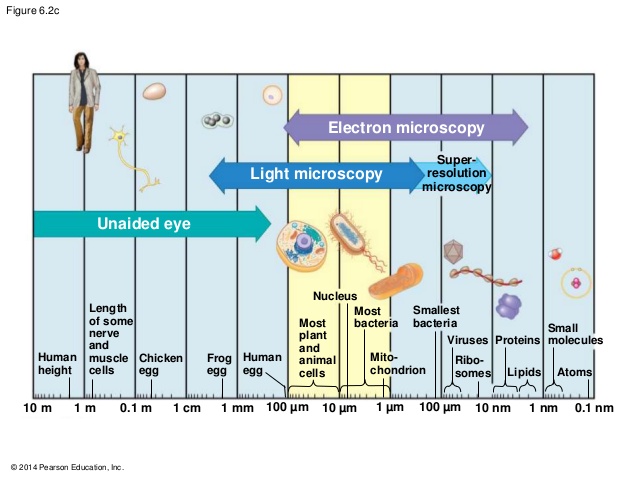
Reference: https://socratic.org/questions/5720d05e7c01493ef1be9590
Light microscope, LM or optical microscope
The optical microscope can observe magnified images using two lenses (objective lens, ocular lens). Optical microscopes are generally divided into a wide variety of different types, depending on the purpose of use, as well as the biological microscopes we use in the laboratory, and have organized the optical microscopes and their characteristics that are mentioned or used relatively often.
Phase-contrast microscope: When light passes through an interval other than a vacuum, phase changes occur through interaction with matter, which in our eyes this phase change is not our eyes. Therefore, it is useful for observing undyeed living cells by converting and amplifying phase changes caused by density differences in specimens into brightness changes.
Fluorescence microscope: Fluoroscopy is a term that encompasses a slightly larger category, and all optical microscopes that are observed by the observer using fluorescence are called fluorescent microscopes. Photographs of fluorescent microscopes, such as genomics, proteomics, or developmental studies, are commonly seen in biological studies because they are often used to locate specific proteins in cells or to observe their pathways over time at the stage of occurrence.
For reference, the green fluorescence protein (GFP), which is commonly used for observing fluorescent microscopes, was originally a protein expressed in jellyfish, but was conveniently developed to enable expression after being introduced regardless of various species. Therefore, studies using GFP fusion proteins are still active today, and three scientists, Roger Y. Tsien, Osamu Shimomura and Martin Chalfie, who discovered and developed them, were awarded the 2008 Nobel Prize for Chemistry in recognition of their contributions.
Confocal Microscope: reconstructs two-dimensional images of cross-section at different depths for the target specimen to produce three-dimensional images.
Deconvolution Microscope: Using deconvolution software, you can reconstruct the blurred images obtained by taking the target samples from different sides to create a clear 3D stereoscopic image.
Electron microscope, EM

Because electron microscope uses electron beam, it has a shorter wavelength than visible light, allowing you to observe the specimen at a higher magnification rate (waveform: 350 to 750 nm of visible light; electron wavelength (1000 kV): 0.00087 nm). However, compared to optical microscopes that can observe living cells, electron microscopes are characterized by more demanding sample processing and only dead samples. The electron microscope is largely divided into transmissive electron microscope and injection electron microscope.
Transmission electron microscope (TEM): TEM uses a technique to image the sensor as the electron passes through the specimen and magnify and observe images of the interaction between the specimen and the electron. Compared to SEM, there is a limitation that samples can be observed at a higher magnification but only in two-dimensional black and white.
Scanning electron microscope (SEM): SEM is to inject electrons into the surface of a sample to detect secondary electrons emitted by atoms excited by the electron wires and to convert them into electrical signals for imaging. Although it has a lower scale than TEM, it has the advantage of being able to observe three-dimensional black and white.References
1. Campbell Biology (10th Edition)
2. WIKIPEDIA
'Biological Science' 카테고리의 다른 글
The invention of a microscope (Hooke and Leeuwenhoek) (0) 2020.02.06 The Nobel Prize in Physiology or Medicine 2019 (0) 2020.02.05